|
 
 |
| ORIGINAL ARTICLE |
|
| Year : 2015 | Volume
: 3
| Issue : 1 | Page : 38-44 |
|
Evaluation of self-etching primer and erbium-doped yttrium aluminum garnet laser on bracket bond strength: An in-vitro study
Pawankumar Dnyandeo Tekale1, Ketan K Vakil2, Jeegar K Vakil3
1 Former Postgraduate Student, Department of Orthodontic, S.M.B.T. Dental College and Hospital, Sangamner, Maharashtra, India
2 Professor and Head, Department of Orthodontic, S.M.B.T. Dental College and Hospital, Sangamner, Maharashtra, India
3 Senior Lecturer, Department of Orthodontic, S.M.B.T. Dental College and Hospital, Sangamner, Maharashtra, India
| Date of Web Publication | 29-Dec-2014 |
Correspondence Address:
Pawankumar Dnyandeo Tekale
Dnyanita Orthodontic Care, Adalat Road, Aurangabad - 431 001, Maharashtra
India
 Source of Support: None, Conflict of Interest: None  | Check |
DOI: 10.4103/2321-3825.146355

Introduction: The purpose of this study was to evaluate and compare the shear bond strength, the adhesive remnant scores and surface characteristics of the teeth prepared for bonding with erbium-doped yttrium aluminum garnet (Er:YAG) hard tissue laser, self-etching primer (SEP) and phosphoric acid etching. Materials and Methods: Seventy-eight human premolars, extracted for orthodontic purposes were randomly divided into three groups, enamel was irradiated with 37% phosphoric acid in Group-1, with SEP in Group-2 and with Er:YAG laser at 1.5-W in Group-3. After surface preparation standard edgewise stainless steel premolar brackets were bonded; one tooth in each group was not bonded and was examined under a scanning electron microscopic. The brackets were debonded 24 h later; shear bond strengths were measured, and adhesive remnant index scores were recorded. Results: Statistically significant differences were found between phosphoric acid etching, SEP, 1.5-W laser irradiation. Adhesive remnant scores were compared with the Chi-square test, and statistically significant differences were found between all groups. Conclusions: The mean shear bond strength obtained with 37% phosphoric acid etching, SEP and Er-YAG laser etching were clinically acceptable. SEP produces a more conservative etch pattern and more shear bond strength than phosphoric acid. More adhesive was left on the SEP treated enamel as compared to enamel treated with acid and laser etching. Keywords: Erbium-doped yttrium aluminium garnet laser, self-etching primer, shear bond
How to cite this article:
Tekale PD, Vakil KK, Vakil JK. Evaluation of self-etching primer and erbium-doped yttrium aluminum garnet laser on bracket bond strength: An in-vitro study. J Orthod Res 2015;3:38-44 |
How to cite this URL:
Tekale PD, Vakil KK, Vakil JK. Evaluation of self-etching primer and erbium-doped yttrium aluminum garnet laser on bracket bond strength: An in-vitro study. J Orthod Res [serial online] 2015 [cited 2018 Sep 6];3:38-44. Available from: http://www.jorthodr.org/text.asp?2015/3/1/38/146355 |
| Introduction | |  |
Efficient orthodontic treatment with fixed appliances requires adequate bonding of brackets to the enamel surfaces of the teeth. Bond failures decrease the efficiency of treatment, resulting in more time in treatment, increased chair time per visit, and greater patient inconvenience. According to the study of Buonocore in 1955, the standard protocol to remove the smear layer for successful bonding has been acid etching. [1] Development of these micromechanical bonds contributes to long-term bonding strength. In 1965, Newman reported the use of epoxy resin for attaching brackets. [2] The bonding procedure improved the overall treatment results by eliminating band occupying interdental spaces, decreased gingival irritation, and easier removal of plaque and decreased risk of calcification. [3] Since then, various adhesives and methods of bonding orthodontic attachments have been reported to enhance the bond strength of the orthodontic attachments. [4],[5] The inimitable properties of new bonding system in operative dentistry is that conjoining conditioning and priming agents into a single acidic primer solution for simultaneous use on both enamel and dentin. [6] Recent advances in dental bonding chemistry allow the combination of the etchant and primer into one product called a self-etch primer (SEP) composed of methacrylated phosphoric acid esters. In the 1990s, SEP was introduced to orthodontics as a way to save chair time during bonding. Bonding practices based on SEPs, originally introduced in restorative dentistry to bond composite resin restorations to both dentin and enamel, are being used in clinical orthodontics. One obvious advantage of using a SEP is to expedite the bonding procedure by combining etching and priming into a single-step. In addition to saving time, fewer steps in the bonding process lead to fewer procedural errors, such as contamination with saliva and water. [7],[8],[9],[10],[11] Lasers are not, however, new to the field; some of the first reports, on in-vitro studies; date back to the late 1960s. It was not until the early 1980s, however that lasers truly saw their first use in clinical practice. When used efficaciously and ethically, lasers are an exceptional modality of treatment for many clinical conditions that dentists or dental specialists treat on a daily basis. The erbium-doped yttrium aluminum garnet (Er:YAG) laser with a wavelength of 2940 nm is highly absorbed by water and hydroxyapatite. Being effective in cutting enamel and dentin, it is the first approved laser tool applied to dental hard tissues in the United States. [12],[13] It was shown that Er:YAG laser-prepared dentin had improved bond strengths when compared with acid-etched groups. [14] However, the tensile strength of bracket-tooth bonds after preparation of the enamel surface by Er:YAG laser etching was inferior to that obtained after conventional acid etching. Nonetheless, if the irradiation parameters can be advertently controlled, the subsurface fissuring that is unfavorable to adhesion can be avoided. [15] Nonetheless, if the irradiation parameters can be advertently controlled, the subsurface fissuring that is unfavorable to adhesion can be avoided.
Until date, there are no reported studies that compare the bond strengths of orthodontic bracket etched by acid, SEP and Er:YAG laser on the enamel surface. The purpose of this study is to assess the shear bond strength and the adhesive remnant index (ARI) scores of teeth prepared for bonding with Er:YAG laser etching, etching with a SEP and etching done with phosphoric acid.
| Materials and Methods | | |
This in-vitro study was done on extracted human premolars to assess the shear bond strength and the ARI scores of teeth prepared for bonding with Er:YAG laser etching, etching with a SEP and etching done with phosphoric acid. Seventy-eight freshly extracted human premolars were collected and stored in a solution of 0.1% (weight/volume) thymol. The inclusion criteria for selection of tooth were the maxillary and mandibular human premolars that the teeth be noncarious, nonhypoplastic, nonfluorosed, healthy with absence of restorations, and no micro-cracks. Stainless steel standard edgewise premolar brackets (3M Unitek) were used for bonding to the tooth surfaces in all the three groups. The bracket base area used for this study was 12.6 mm 2 as given by the manufacturer.
The 78 teeth were randomly divided into three groups. 75 teeth were embedded in self-cure acrylic resin in mold. 75 teeth were done with shear bond testing and ARI evaluation. One tooth from each group was taken for scanning electron microscope (SEM) evaluation to examine the etching pattern. Three teeth which were prepared for SEM evaluation were not embedded in acrylic resin.
The groups were divided as follows:
- Group-1 (control): Conventional acid etching technique (n = 25)
- Group-2: Etching with SEP (n = 25)
- Group-3: Etching with laser (Er:YAG) (n = 25).
All the teeth were polished with a polishing paste (fluoride-free and oil-free) to remove any residual plaque or stains by using a contra-angle micromotor handpiece and a polishing brush.
Group-1: On 25 teeth, 37% phosphoric acid gel was applied to the enamel surface to be bonded for 15-s, followed by a 10-s water rinse and a 5-s moisture free and oil-free air dry. The sealant was applied, and the brackets were bonded with Transbond XT (3M Unitek, Monrovia, California) and light cured for 40-s according to the manufacturer's instructions.
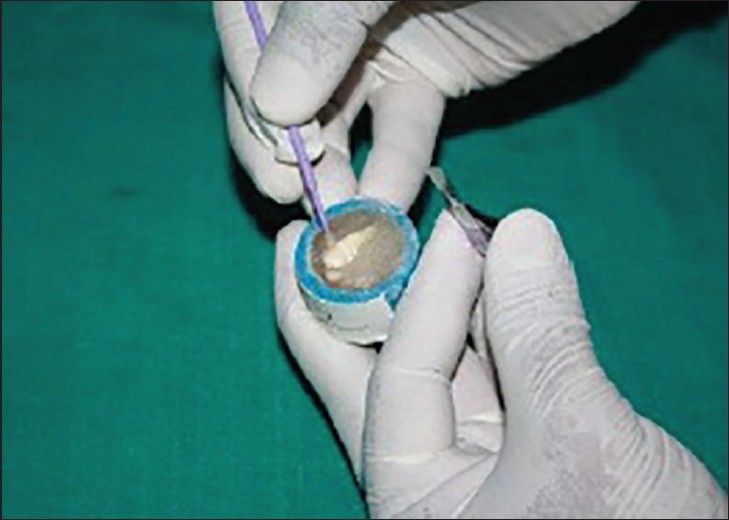
Group-2: On 25 teeth, Transbond SEP was applied for 5-s as single layer with applicator sponge provided by manufacture and slightly air dried (5-s) as per manufacturer's instruction. The sealant was applied, and the brackets were bonded with Transbond XT (3M Unitek) and light cured for 40-s according to the manufacturer's instructions [Figure 1].
Group-3: On 25 teeth, enamel was etched with Er:YAG laser with 1.5-W for 15-s as per manufacturer's instruction. After etching, the sealant was applied, and the brackets were bonded with Transbond XT (3M Unitek) and light cured for 40-s according to the manufacturer's instructions [Figure 2].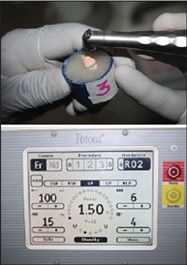 | Figure 2: Etching with erbium-doped yttrium aluminum garnet laser for Group-3 sample
Click here to view |
The specimens were stored in water at 37°C for 24 h in distilled water.
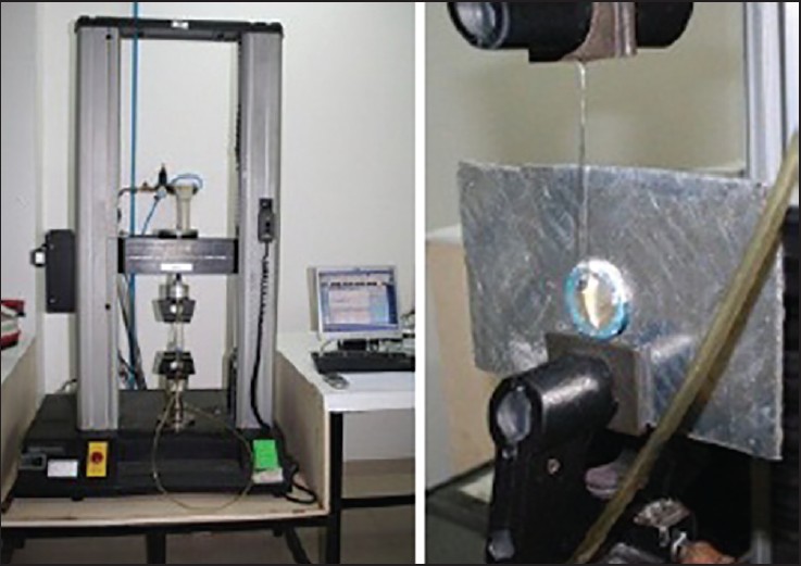
Shear bond strength test was done with an Instron Universal Testing (Instron model no. 33R 4467, Instron 182 Ltd., Buckinghamshire, England) [Figure 3]. The machine was set and calibrated according to manufacturer's instructions. The specimens were secured in lower jaws of machine so that bonded bracket bases were parallel to the shear force direction.
The samples were tested for shear bond strength on Instron Universal Testing machine at a cross head speed of 2.5 mm/min. The maximum load (shear bond strength) necessary to debond the bracket was recorded in Newtons and then converted into Megapascals as a ratio of Newtons to surface area of the bracket (MPa = N/mm 2 ).
Once the brackets were debonded, the enamel surface of each tooth was examined and the ARI scores were recorded under stereomicroscope (Magnum, Olympus India Pvt. Ltd., New Delhi) at ×10 magnification to evaluate the amount of resin remaining on the tooth. The ARI [16] was used to describe the quantity of resin remaining on the tooth surfaces. These scores were as follows: 0, no adhesive remained on the tooth;
- Less than half of the enamel bonding site was covered with adhesive;
- More than half of the enamel bonding site was covered with adhesive; and
- The enamel bonding site was covered entirely with adhesive.
For the three samples (one from each group), which were not embedded in acrylic resin, to assess the etching efficacy of intact enamel, using a SEM (JEOL JSM 6360, Germany), buccal enamel surfaces were conditioned with same enamel surface etching protocol that were followed in Group 1, 2, and 3 just prior to sputtering. After conditioning or etching, the specimens were sputter-coated with platinum (JEOL JSM 6360, Germany) and examined by a SEM operating at 10 kV for evaluation of the different etching pattern.
Statistical Analysis
The statistical software SPSS for Windows, version 10.0.0, SPSS, Chicago, lll was used for the analysis of the data. Descriptive statistical analysis was carried out in the present study. Descriptive statistics, including the mean, standard deviation, maximum and minimum shear bond strength values were calculated for each of the three groups. The comparisons between the three groups were made by ANOVA test. To assess the difference in the shear bond strength in between groups, Independent sample t-test was done. The Chi-square test was used to evaluate differences in ARI scores between the groups.
| Results | | |
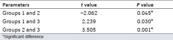
The bond strength, for Group-1 (37% phosphoric acid etching), Group-2 (etching with SEP) and Group-3 (Er:YAG Laser with 1.5-W) were recorded. The mean shear bond strength, standard deviation and standard error of mean among three groups presented in [Table 1]. The comparisons between the three groups were made and P value for bond strength was derived by ANOVA test, presented in [Table 2]. To assess the difference in the shear bond strength in between groups, Independent sample t-test was done, presented in [Table 3]. Graph 1 represents the mean bond strength and mean peak load among the three groups.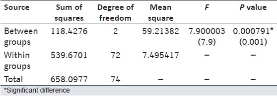 | Table 2: Comparisons between the three groups and P value for bond strength derived by ANOVA test
Click here to view |
 | Table 3: Comparisons between the three groups and P value for bond strength derived by Independent sample t-test
Click here to view |
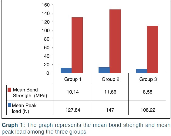
The results indicated that the mean shear bond strengths obtained with 37% phosphoric acid, SEP and Er:YAG laser were 10.14, 11.66 and 8.58 MPa respectively. When the acid etch and SEPs were compared, the P = 0.045 indicating that there was significant difference in the bond strength between these groups. Similarly, it was found that there was significant difference in the bond strength between the acid etch group and the laser etch group (P = 0.030) and between SEP and the laser etch group (P = 0.001).
Adhesive Remnant Index Evaluation
On comparison of the ARI scores, it was found that the adhesive left on the enamel surface after debonding was significantly higher in the SEP and laser etched group than the acid-etched group. There was significant difference in the ARI scores between the acid etch group and the SEP; the acid etch group and laser etch group; the self-etch group and laser etch group. The value for ARI score was recorded and P value is calculated by Chi-square test was described in [Table 4]. Graph 1 represented the mean ARI scores among the three groups.
Scanning Electron Microscope Examination
For the three samples (one from each group) which were not embedded in acrylic resin, to assess the etching efficacy of intact enamel, using a SEM (JEOL JSM 6360, Germany), buccal enamel surfaces of the crown of the teeth were conditioned with same enamel surface etching protocol that was followed in Groups 1, 2 and 3 just prior to sputtering. After conditioning or etching, the specimens were sputter-coated with platinum (JEOL JSM 6360, Germany) and examined by SEM operating at 10 kV.
Scanning electron microscope evaluations of the samples showed some different surface characteristics.
Group-1 shows an enamel surface with a type 1 etching pattern. There was generalized roughening of the enamel surface, but with a distinct pattern showing hollowing of prism centers with relatively intact peripheral regions, as described by Silverstone et al. [17] The hydroxyapatite dissolved by phosphoric acid produced tags and rough surfaces that provided the mechanical inter-locking for the resin [Figure 4].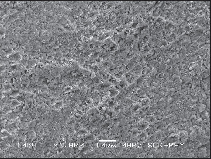 | Figure 4: Scanning electron microscopic image of Group-1 sample after etching with 37% phosphoric acid
Click here to view |
Group-2 shows an enamel surface with a type 2 etching pattern. The prism peripheries appeared to be removed and the prism cores were left projecting towards the original surface, as described by Silverstone et al. [17] The use of Transbond Plus SEP produced a uniform etch pattern that was more conservative and less destructive to the enamel surface. Consequently, a regular resin tag distribution was observed, which showed less magnitude when compared with the acid group as described in previous study [18] [Figure 5].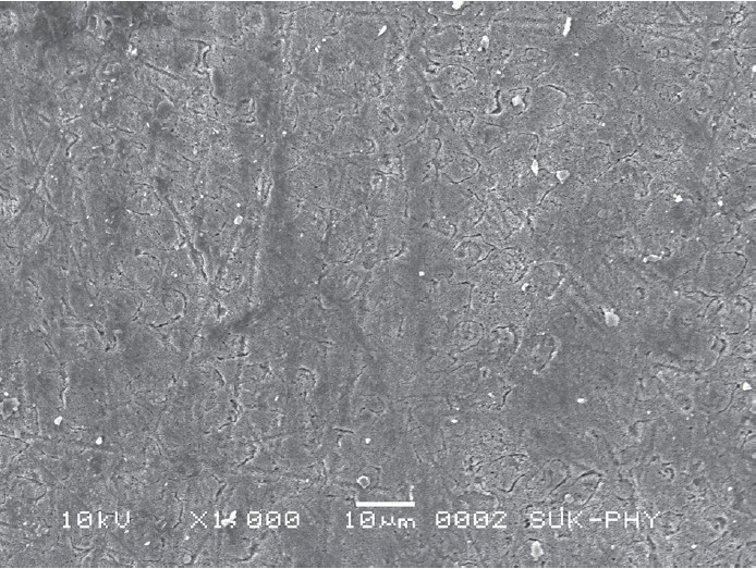 | Figure 5: Scanning electron microscopic image of Group-2 sample after etching with self-etching primer
Click here to view |
In Group-3, laser irradiation with 1.5-W produced type 3 etching pattern. The area appeared as a generalized surface roughening with regions resembling hollowed prism centers adjacent to the area in which the pattern was more consistent with the loss of prism peripheries, as described by Silverstone et al. [17] The laser-ablated surfaces were accompanied by the appearance of micro-cracks that aid the penetration of resin as described in previous study [19] [Figure 6].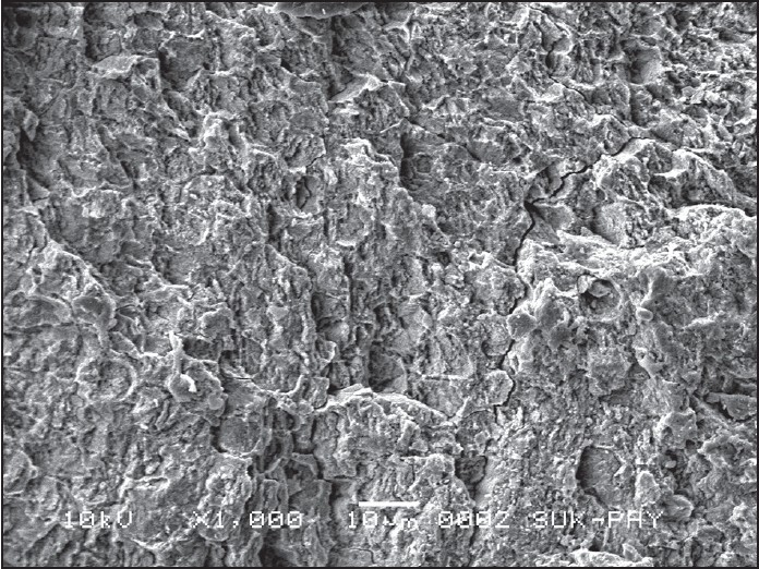 | Figure 6: Scanning electron microscopic image of Group-3 sample after etching with erbium-doped yttrium aluminum garnet laser at 1.5-W power
Click here to view |
| Discussion | | |
The potential disadvantage of enamel acid etching is the demineralization of the most superficial layer and the development of white-spot lesions around bonded orthodontic appliances, as a result of demineralization, the surface becomes more susceptible to long-term acid attack and caries, especially when resin impregnation is defective because of air bubbles or saliva contamination. These effects are particularly important because plaque tends to accumulate adjacent to the bonded orthodontic attachments. [1],[12],[20] It was estimated that saliva contamination reduces bond strength because it fills the microporosities of the etched enamel, affecting the mechanical retention of the bonding material. [7]
The continuing developments in dental material science have led to improvements in adhesive bonding formulations, resulting in the current availability of a wide range of products, including the single-step etch/primer solutions. In a SEP, the active ingredient is a methacrylated phosphoric acid ester. The phosphoric acid and the methacrylate group are combined into a molecule that etches and primes at the same time. The phosphate group on the methacrylated phosphoric acid ester dissolves the calcium and removes it from the hydroxyapatite. However rather than being rinsed away, the calcium forms a complex with the phosphate group and gets incorporated into the network when the primer polymerizes. Agitating the primer on the tooth surface serves to ensure that fresh primer is transported to the enamel surface. Etching and monomer penetration to the exposed enamel rods are simultaneous. In this manner, the depth of the etch is identical to that of the primer penetration. Three mechanisms act to stop the etching process. First, the acid groups attached to the etching monomer are neutralized in a similar way, as is phosphoric acid, by forming a complex with the calcium from the hydroxyapatite. Second, as the solvent is driven from the primer during the airburst step, the viscosity rises, slowing the transport of acid groups to the enamel interface. Finally, as the primer is light cured and the primer monomers are polymerized, transport of acid groups to the interface is stopped. [21],[22] If bond failure rates are comparable with those after the use of conventional acid etching, then the additional time saved at the beginning of treatment should make the SEP more cost-effective. [23]
For many years, the Er:YAG laser has been applied in dentistry for carious lesion removal, cavity preparation, endodontic procedures, and surface conditioning. The Er:YAG laser is able to handle dental hard tissue with high efficiency because of the high absorbability of the 2.94 μm wavelength by water and dental enamel. The absorbed laser energy is converted to heat that boils water in the tooth, forming high-pressure steam. The successive explosive vaporization of water modifies the smooth tooth surface, creating an irregularly serrated and microfissured morphology. [12],[14]
Etching of enamel surface with an Er:YAG laser system yielded clinically acceptable and similar bond strengths to acid etching with 37% orthophosphoric acid for 15-30 s. In addition, laser-induced caries resistance would also be of great importance in orthodontics. Furthermore, lasers might save some clinical time. [12]
The present study evaluated the shear bond strength obtained with conventional 37% phosphoric acid etching procedure with the SEP (Transbond Plus, 3M Unitek, Monrovia, California) etching and use of Er-YAG laser etching. The results indicated that the mean shear bond strengths obtained with 37% phosphoric acid, SEP and Er:YAG laser were 10.14, 11.66 and 8.58 MPa, respectively. Clinically, acceptable bond strengths have been reported to range from 6 to 8 MPa. [5] In this experiment, mean bond strength of all groups was well above this minimal requirement. These bond strengths are considered to be able to withstand masticatory and orthodontic forces.
When the Groups 1 and 2 were compared in the ANOVA test there was significant difference in the bond strength between these groups. Further evaluation with Independent sample t-test between Groups 1 and 2, the P = 0.045 and t = −2.062, indicating the significant difference. The negative value of t indicates that the shear strength of teeth treated with Transbond SEP is 1.52 units stronger than teeth etched with phosphoric acid.
When Groups 1 and 3 were analyzed with the ANOVA test, significant difference was found. Further evaluation with Independent sample t-test between Groups 1 and 3, the P = 0.030, which was <0.05 indicating the significant difference between the Groups 1 and 3. Though the mean shear bond strength value of the laser etched group was less than acid etch group, it had clinically acceptable value.
On statistical analysis with ANOVA test, the significant difference was found between the Groups 2 and 3. Further evaluation with Independent sample t-test, P = 0.001 indicating the statistically significant difference among Groups 2 and 3. Group 2 had mean shear bond strength more than Group-3 and both were in clinical acceptable range.
The evaluation of ARI scores showed statistically significant difference among acid etch group, self-etch and laser etched group. The ARI score was significantly high in the self-etch group as compared to the acid etch and laser etch group. This could be an advantage or a disadvantage. Less chair time is needed with less adhesive left on the enamel after debonding, but it might cause enamel fracture, while debonding, especially with ceramic brackets.
In a study done by Hosein et al. on enamel loss during bonding, debonding, and cleanup with use of a SEP, it was found that the enamel surface treatment with the conventional acid-etching technique leads to more enamel loss, leaves more adhesive on enamel after debonding and there is more enamel loss at enamel clean up than with the use of a SEP. [24] Hence, in addition to saving clinical time during bonding, the SEPs have also been reported to save clinical time at the time of debonding and clean up. It was also found that laser at 1.5-W power produced clinically acceptable shear bond strength which was comparable to the levels achieved with conventional 37% phosphoric acid etching. Moreover, Er:YAG laser etching is painless and does not involve either vibration or heat, and the easy handling of the apparatus makes this treatment highly attractive for routine clinical use. [25] Furthermore, this laser can be used in wet conditions and the water-cooled system does not cause any untoward thermal effects on the tooth pulp. Furthermore, the clinician has more control of the area to be etched with the laser system. Although gel acids are more stable than liquid acids, there is always a spread of acid on the enamel surface. In addition, laser-induced caries resistance would also be of great importance in orthodontics. In some previous studies, the recommendation of SEPs and Lasers for orthodontic bonding is evident. [8],[9],[17],[18]
The result showed that all the three procedure produced clinically acceptable bond strength. However, this was an in-vitro study and the results may vary when procedure are actually done on patient. Hence further in-vivo studies are suggested.
| Summary and Conclusion | | |
Under the conditions of this in-vitro study, it was found the following:
- The results indicated that the mean shear bond strengths obtained with 37% phosphoric acid, SEP and Er:YAG laser were 10.14, 11.66 and 8.58 MPa respectively. The bond strength values in this study were clinically acceptable.
- There was significant difference in the bond strength obtained with 37% phosphoric acid etch group, SEP group and the Er:YAG laser irradiated group.
- The mean shear strength of teeth treated with Transbond SEP is 1.52 units stronger than teeth etched with phosphoric acid.
- The mean shear bond strength obtained with Er-YAG laser etching procedure was less than mean shear bond strength obtained with 37% phosphoric acid etching, but bond strength values of Er-YAG laser etching were clinically acceptable.
- More adhesive was left on the SEP treated enamel when compared to enamel treated with acid and laser etching.
- SEP produces a more conservative etch pattern and more shear bond strength than phosphoric acid, thereby minimizing the enamel loss, was confirmed in this study.
| References | | |
| 1. | Buonocore MG. A simple method of increasing the adhesion of acrylic filling materials to enamel surfaces. J Dent Res 1955;34:849-53.  [ PUBMED] |
| 2. | Newman GV. Epoxy adhesives for orthodontic attachments: Progress report. Am J Orthod 1965;51:901-12. [ PUBMED] |
| 3. | Newman GV, Snyder WH, Wilson CE Jr. Acrylic adhesives for bonding attachments to tooth surfaces. Angle Orthod 1968;38:12-8. |
| 4. | Retief DH, Dreyer CJ, Gavron G. The direct bonding of orthodontic attachments to teeth by means of an epoxy resin adhesive. Am J Orthod 1970;58:21-40. [ PUBMED] |
| 5. | Reynolds JR. A review of direct bonding. Br J Orthod 1975;2:171-80. |
| 6. | Chigira H, Koike T, Hasegawa T, Itoh K, Wakumoto S, Hayakawa T. Effect of the self etching dentin primers on the bonding efficacy of a dentin adhesive. Dent Mater J 1989;8: 86-92. [ PUBMED] |
| 7. | Paschos E, Westphal JO, Ilie N, Huth KC, Hickel R, Rudzki-Janson I. Artificial saliva contamination effects on bond strength of self-etching primers. Angle Orthod 2008;78:716-21. |
| 8. | Bishara SE, Otsby AW, Ajlouni R, Laffoon J, Warren JJ. A new premixed self-etch adhesive for bonding orthodontic brackets. Angle Orthod 2008;78:1101-4. |
| 9. | Vilchis RJ, Hotta Y, Yamamoto K. Examination of enamel-adhesive interface with focused ion beam and scanning electron microscopy. Am J Orthod Dentofacial Orthop 2007;131:646-50. |
| 10. | Eliades T. Orthodontic materials research and applications: Part 1. Current status and projected future developments in bonding and adhesives. Am J Orthod Dentofacial Orthop 2006;130:445-51. |
| 11. | Vicente A, Bravo LA, Romero M. Influence of a nonrinse conditioner on the bond strength of brackets bonded with a resin adhesive system. Angle Orthod 2005;75:400-5. |
| 12. | Hibst R, Keller U. Experimental studies of the application of the Er:YAG laser on dental hard substances: I. Measurement of the ablation rate. Lasers Surg Med 1989;9:338-44. |
| 13. | Li ZZ, Code JE, Van De Merwe WP. Er:YAG laser ablation of enamel and dentin of human teeth: Determination of ablation rates at various fluences and pulse repetition rates. Lasers Surg Med 1992;12:625-30. |
| 14. | Visuri SR, Gilbert JL, Wright DD, Wigdor HA, Walsh JT Jr. Shear strength of composite bonded to Er:YAG laser-prepared dentin. J Dent Res 1996;75:599-605. |
| 15. | Martínez-Insua A, Da Silva Dominguez L, Rivera FG, Santana-Penín UA. Differences in bonding to acid-etched or Er:YAG-laser-treated enamel and dentin surfaces. J Prosthet Dent 2000;84:280-8. |
| 16. | Artun J, Bergland S. Clinical trials with crystal growth conditioning as an alternative to acid-etch enamel pretreatment. Am J Orthod 1984;85:333-40. [ PUBMED] |
| 17. | Silverstone LM, Saxton CA, Dogon IL, Fejerskov O. Variation in the pattern of acid etching of human dental enamel examined by scanning electron microscopy. Caries Res 1975;9:373-87. [ PUBMED] |
| 18. | Lee BS, Hsieh TT, Lee YL, Lan WH, Hsu YJ, Wen PH, et al. Bond strengths of orthodontic bracket after acid-etched, Er:YAG laser-irradiated and combined treatment on enamel surface. Angle Orthod 2003;73:565-70. |
| 19. | Cal-Neto JP, Miguel JA. Scanning electron microscopy evaluation of the bonding mechanism of a self-etching primer on enamel. Angle Orthod 2006;76:132-6. |
| 20. | Maijer R, Smith DC. Crystal growth on the outer enamel surface - An alternative to acid etching. Am J Orthod 1986;89:183-93. [ PUBMED] |
| 21. | Aljubouri YD, Millett DT, Gilmour WH. Laboratory evaluation of a self-etching primer for orthodontic bonding. Eur J Orthod 2003;25:411-5. |
| 22. | Buyukyilmaz T, Usumez S, Karaman AI. Effect of self-etching primers on bond strength - Are they reliable? Angle Orthod 2003;73:64-70. |
| 23. | Bishara SE, VonWald L, Laffoon JF, Warren JJ. Effect of a self-etch primer/adhesive on the shear bond strength of orthodontic brackets. Am J Orthod Dentofacial Orthop 2001;119:621-4. |
| 24. | Hosein I, Sherriff M, Ireland AJ. Enamel loss during bonding, debonding, and cleanup with use of a self-etching primer. Am J Orthod Dentofacial Orthop 2004;126:717-24. |
| 25. | Ozer T, Baºaran G, Berk N. Laser etching of enamel for orthodontic bonding. Am J Orthod Dentofacial Orthop 2008;134:193-7. |
[Figure 1], [Figure 2], [Figure 3], [Figure 4], [Figure 5], [Figure 6]
[Table 1], [Table 2], [Table 3], [Table 4]
|


 Search Pubmed for