|


 |
| CASE REPORT |
|
| Year : 2013 | Volume
: 5
| Issue : 2 | Page : 89-91 |
|
|
Congenital uterovaginal prolapse present at birth
Ekwunife Okechukwu Hyginus1, Chukwuka Onuora John2
1 Department of Surgery, Nnamdi Azikiwe University Teaching Hospital, Nnewi Anambra, Nigeria
2 Department of Paediatrics, Nnamdi Azikiwe University Teaching Hospital, Nnewi Anambra, Nigeria
| Date of Web Publication | 13-Mar-2014 |
Correspondence Address:
Ekwunife Okechukwu Hyginus
Departments of Surgery, Nnamdi Azikiwe University Teaching Hospital, Nnewi, Anambra
Nigeria
 Source of Support: None, Conflict of Interest: None
DOI: 10.4103/2006-8808.128741

 Abstract Abstract | | |
Uterovaginal prolapse presenting at birth is very rare. The cause is attributed to conditions that can cause poor innervation or weakness of the pelvic floor muscle and the supporting ligaments. Different methods of treatment have been used in the past to reduce and maintain reduction of the prolapse. We report a case of a congenital UVP in a day old child noticed at delivery. He was delivered breech and had a sacral dimple with a tuft of hair. He was successfully managed conservatively with digital reduction and strapping of the buttocks down to the legs with crepe bandage for 72 h. Keywords: Breech, congenital, Nigeria, prolapse, spina bifida, utero-vaginal
How to cite this article:
Hyginus EO, John CO. Congenital uterovaginal prolapse present at birth. J Surg Tech Case Report 2013;5:89-91 |
| Introduction | |  |
Uterovaginal prolapse (UVP) presenting at birth is very rare. To the best of our knowledge, not more than 20 cases of neonatal UVP have been reported in English literature. [1],[2],[3],[4],[5],[6],[7],[8],[9],[10],[11] Utero-vaginal prolapse is the downward descent and protrusion of the uterus and vagina to the exterior via the introitus. It is as a result of weakness of the cardinal ligaments and uterosacral ligaments which provide support to the uterus. Most reported neonatal UVP were either 3 rd degree or procidentia. [1],[2],[3],[4],[5],[6],[7],[8],[9],[10],[11] This is a report of a congenital UVP (3 rd degree) in a 12 h-old-child noticed at delivery.
| Case Report | | |
A 12-hour-old female neonate was presented to us with a fleshy mass protruding from the vulva since birth. The baby is a product of term gestation and was delivered breech. Labor was not prolonged, and there was no instrumental delivery. The baby passed meconium on the 1 st day of birth and tolerated breast milk feeding.
The mother, a 31-year-old, para 3, seamstress had recurrent febrile conditions throughout the pregnancy and was treated at the primary health center where she booked. She neither ingested herbal medications nor any unprescribed drugs during pregnancy.
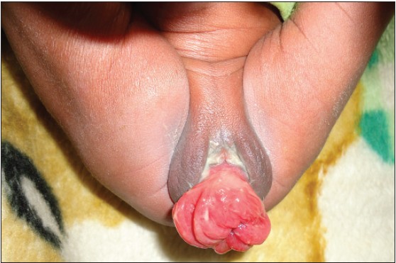
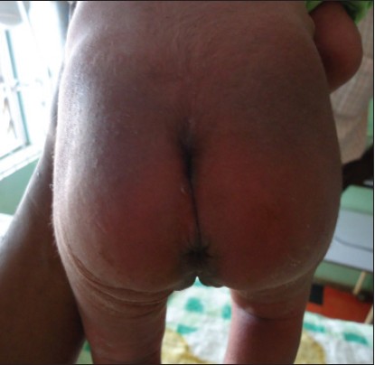
On examination, the child weighed 3.0 kg. There was a fleshy, reddish, and edematous mass protruding from the vulva [Figure 1]. The wall was thrown into folds. At the apex there was a blind-ended opening. The urethral opening was located separately above the mass. The anus was patent, and meconium stained finger noted on digital rectal examination. She has a sacral dimple with tufts of hair in it [Figure 2]. Lower limb motor activity, sensations, respiratory, and cardiovascular systems were all normal. No masses were felt on abdominal examination. The skin was neither wrinkled nor thrown into folds. Skin elasticity was normal.
Under sedation, size 6 Foley's urethral catheter was passed into the bladder. The mass was reduced by gripping it with the right hand and gently pushing it inwards. To prevent re-protrusion on straining, both lower limbs were strapped together using crepe bandage which was applied in a mermaid fashion extending from the buttocks to the lower legs sparing the anus for defecation. Both catheter and bandaging were removed after 72 h. Following successful reduction, the parents were unwilling to neither subject the child to further investigations nor comply with follow-up schedules. However, through contact tracing, the prolapse has not recurred 6 months after, and the child is growing appropriately.
| Discussion | | |
The uterus and the vagina are essentially supported by the muscular pelvic diaphragm and the three condensations of the endopelvic fascia (cardinal ligaments, uterosacral ligaments, and pubocervical fascia). [12]
Congenital UVP usually results from the weakness of the pelvic muscular support and the ligaments. This may be secondary to either congenital weakness in the pelvic musculature or defects in innervation. In adults the uterovaginal junction is angulated with the uterus lying almost horizontal to the muscular pelvic diaphragm. In the fetus and the neonate there is minimal or no angulation and the orientation is almost vertical, as the true pelvis is not well-formed yet and the pelvic organs are essentially abdominal. Hence, raised intra-abdominal pressure as occurs during prolonged breech delivery is transmitted straight down the uterus and out through the vagina and introitus. This becomes pronounced if the pelvic muscle is weak and the ligaments stretched.
However, the etiology of congenital UVP is not well-established. Most of the few reported cases were often associated with some identifiable risk factors [Table 1]. Spina bifida, especially myelomeningocele is the most common risk factor (85% of cases). [1],[2],[3],[4] Occult spina bifida as occurred in the index patient is not common.
Defective innervations of the pelvic floor muscle in meningomyelocele may lead to weakened support of the uterus and vagina. The rise in fetal intra-abdominal pressure during breech delivery stretches the pelvic floor muscle and the ligaments thus increasing the risk of prolapse. Our patient had features of occult spina bifida and was delivered breech. Congenital UVP is also seen in neonates with congenital cutis laxa. [5] This is a genetic disorder characterized by generalized loose and redundant skin with reduced elasticity. Inheritance can be autosomal dominant, autosomal recessive, or X-linked. [13] The babies present with loose lax skin with reduced elastic recoil. Hernias and rectal prolapse are common associations in this condition. The index patient did not clinically manifest any skin abnormality.
Early treatment of prolapse is important to prevent injury and metaplasia of the endometrial lining from prolonged exposure. This possibly may affect fertility later in life. Several modalities which can be either conservative or operative have been used in the treatment of genital prolapse in the neonates. Most authors favor initial digital reduction and conservative management. [1],[6],[9],[10] From the reviewed case reports, conservative management has a success rate of more than 90%, though long-term outcome could not be ascertained yet. The major challenge after the initial digital reduction is the recurrence of prolapse once the intra-abdominal pressure rises during crying or straining. Different authors have overtime evolved different methods of solving this. These methods include the use of various forms of pessaries, [1],[14] vaginal tampons, [6] use of a two-way catheter partially inflated in the vagina to prevent recurrence of reduced prolapse, [9] purse string suturing of vaginal wall with 2-0 catgut, [4] and even temporary fusion of the labia. [11] Baskaran et al., reported a successful outcome with purse string suturing of the vaginal wall in two rows in a neonate with myelomeningocele. [4]
With the resolution of the edema, reduction in the maternal derived estrogen level, the uterus becomes fixed in the normal position in most infants. However, the use of foreign agents can be complicated with cervicitis and vaginitis. [9] Surgical procedures like uterine ventrosuspension, sling, sacral cervicopexy, or abdominal sacrocolpopexy have also been used in recalcitrant cases. [7]
In our patient, re-protrusion occurred immediately after digital reduction. We solved this by strapping the buttocks together by applying crepe bandage from the lower abdomen down to the lower third of the legs in a "mermaid" fashion, sparing the anus for defecation. This was removed after 3 days with no further recurrence. This method has the advantage of simplicity and easy applicability by even the labor attendants at the primary centers soon after the prolapse is observed, before edema and mucosal ulceration sets in.
In conclusion, UVP presenting at birth is a rare condition. The possible risk factors present in this patient include breech delivery and spina bifida occulta. Successful reduction was achieved by digital reduction and strapping the buttocks together with crepe bandage in a "mermaid" fashion.
| References | | |
| 1. | Lockwood G, Durkee C, Groth T. Genital prolapse causing urinary obstruction and hydronephrosis in a neonate: A case and review of the literature. J Neonat Surg 2012;1:39. 
|
| 2. | Taksande AM, Vilhekar KY, Batra P, Jain M. Neonatal genital prolapse. J Indian Med Assoc 2011;109:502-3.
|
| 3. | Mehta MH, Patel RV, Bhatt YC, Mehta L. Neonatal uterine prolapse. Indian J Pediatr 1992;59:644-5.
[PUBMED] |
| 4. | Baskaran D, Mohan P, Nazeeb. Purse string suturing in a neonatal prolapsed uterus. Indian J Surg 2012;74:143-5.
|
| 5. | Choudhary SV, Bisati S, Koley S. Congenital cutis laxa with rectal and utero-vaginal prolapse. Indian J Dermatol Venereol Leprol 2011;77:321-4.
[PUBMED]  |
| 6. | Ellis JB, Boes EG. Genital prolapse. S Afr Med J 1986;69:836.
[PUBMED] |
| 7. | Pirgon O, Atabek ME, Suleymanoglu S. Genital prolapse in a newborn following resection of sacrococcygeal teratoma. J Pediatr Adolesc Gynecol 2009;22:96-8.
|
| 8. | Bader D, Davidovitch M, Berger A. Genital prolapse in a preterm female infant. J Perinatol 1993;13:159-61.
|
| 9. | Abdelsalam SE, Desouki NM, Abd alaal NA. Use of Foley catheter for management of neonatal genital prolapse: Case report and review of the literature. J Pediatr Surg 2006;41:449-52.
|
| 10. | Shuwarger D, Young RL. Management of neonatal genital prolapse: Case reports and historic review. Obstet Gynecol 1985;66:61-3.
|
| 11. | Ajabor LN, Okojie SE. Genital prolapse in the newborn. Int Surg 1976;61:496-7.
[PUBMED] |
| 12. | Ellis H. Clinical Anatomy A revision and applied anatomy for clinical students. 11 th ed. Massachusetts: Blackwell Publishing; 2006. p. 136-49.
|
| 13. | Hucthagowder V, Sausgruber N, Kim KH, Angle B, Marmorstein LY, Urban Z. Fibulin-4: A novel gene for an autosomal recessive cutis laxa. Am J Hum Genet 2006;78:1075-80.
|
| 14. | Loret de Mola JR, Carpentar SE. Management of genital prolapse in neonate and young women. Obstet Gynecol Surv 1996;51:253-60.
|
[Figure 1], [Figure 2]
[Table 1]
|