|


 |
| CASE REPORT |
|
| Year : 2013 | Volume
: 5
| Issue : 2 | Page : 85-88 |
|
|
Dentigerous cyst associated with ectopic canine and a supernumerary tooth: A rare occurrence
Ashwini Ramakrishna1, Pravin Lambade2
1 Department of Oral Pathology and Microbiology, College of Dental Sciences, Davangere, Karnataka, India
2 Department of Oral and Maxillofacial Surgery, Swargiya Dadasaheb Kalmegh Smruti Dental College and Hospital, Nagpur, Maharashtra, India
| Date of Web Publication | 13-Mar-2014 |
Correspondence Address:
Pravin Lambade
Department of Oral and Maxillofacial Surgery, Swargiya Dadasaheb Kalmegh Smruti Dental College and Hospital, Nagpur 441 110, Maharashtra
India
 Source of Support: None, Conflict of Interest: None
DOI: 10.4103/2006-8808.128738

 Abstract Abstract | | |
Amongst the cysts of the jaw dentigerous cyst (DC) is one of the most prevalent types of odontogenic cysts, which is associated with the crown of an unerupted or developing tooth. DC is more commonly seen with mandibular third molar and maxillary canine and rarely other teeth are involved. These cysts seldom associate with supernumerary teeth. The purpose of this article is to describe a case of large dentigerous cyst associated with supernumerary teeth and an ectopic canine, which is a rare presentation along with its management. Keywords: Dentigerous cyst, ectopic teeth, supernumerary teeth
How to cite this article:
Ramakrishna A, Lambade P. Dentigerous cyst associated with ectopic canine and a supernumerary tooth: A rare occurrence. J Surg Tech Case Report 2013;5:85-8 |
How to cite this URL:
Ramakrishna A, Lambade P. Dentigerous cyst associated with ectopic canine and a supernumerary tooth: A rare occurrence. J Surg Tech Case Report [serial online] 2013 [cited 2016 May 25];5:85-8. Available from: http://www.jstcr.org/text.asp?2013/5/2/85/128738 |
| Introduction | |  |
Amongst the developmental cysts of the maxillofacial region dentigerous cyst (DC) is the most common. [1] The cystic lining is derived from the epithelial remanants of tooth forming organ. [2] Second and third decade of life is the common age of occurrence and generally involves the mandibular third molar, maxillary canines and mandibular premolars, followed by supernumerary teeth and central incisor in decreasing order of frequency of involvement. [3]
Those teeth located in the jawbones or in regions other than the alveolar arch are said to be ectopically placed. This may be due to irregularity in the migration of a tooth bud which occurs due to genetic/environmental relationship factors causing a budding tooth to congenitally migrate in the initial stages of embryogenesis, or is the result of displacement of the teeth owing to local factors. Volumetric incompatibility between the tooth and dental arch, prolonged retention of primary teeth, presence of clefts, ankylosis, cystic or neoplastic lesion or trauma may be the local factors involved. [4] In the permanent dentition, the maxillary canine has the longest path of eruption and this may be an etiology for ectopic placement. [5] Supernumerary teeth are uncommon, can be ectopically placed and seldom associate with dentigerous cysts.
| Case Report | | |
A 10 year-old boy presented with a painless swelling in the left maxillary anterior region of 8-9 months duration. On general examination, the patient was apparently healthy with normal growth and development for his age and there was no history of systemic diseases or trauma. Clinical examination revealed an extraoral swelling on the left side of the face with elevation of the nostril and lateral border of the nose. The swelling was extending from the lateral wall of the nose to zygomatic region mediolaterally and from infraorbital region to right upper lip superoinferiorly. There was obliteration of the nasolabial fold. The swelling was soft, fluctuant, painless with positive crepitus.
Intraorally the swelling was extending from maxillary left central incisor to the premolar region with obliteration of the vestibule [Figure 1]. The mucosa overlying the swelling appeared to be intact. Paranasal view of the radiograph [Figure 2] showed a well defined unilocular radiolucency surrounding two teeth in the left anterior region of the maxilla. Displacement of the roots of the maxillary left lateral incisor was also seen. The lesion was extending from the lateral wall of the nose to zygomatico maxillary region mediolaterally and from maxillary alveolar ridge to infraorbital rim superoinferiorly.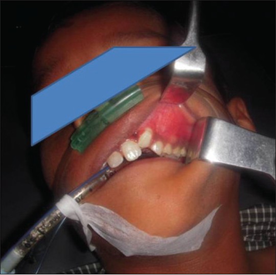 | Figure 1: Pre-operative clinical picture showing swelling in the left maxillary anterior region
Click here to view |
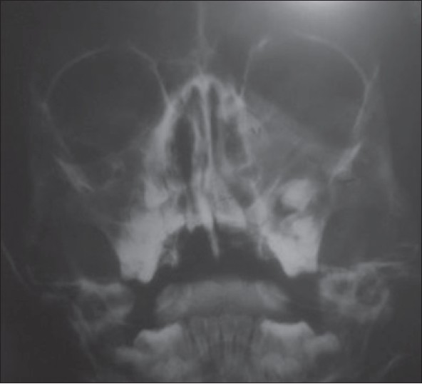 | Figure 2: Paranasal sinus view showing well defi ned unilocular radiolucency surrounding the canine and supernumerary teeth
Click here to view |
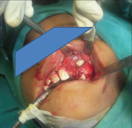
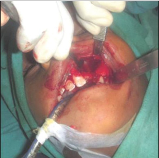
Fine needle aspiration revealed the presence of straw colored fluid within the lesion. After clinical and radiographic examination, a provisional diagnosis of dentigerous cyst involving impacted teeth was made. Routine hematological investigations was normal. Surgical excision of the lesion was planned under general anesthesia. All aseptic precautions were taken. Crestal incision was given in the region of left maxillary central incisor and extended as crevicular incision around left maxillary first premolar. Vertical releasing incision was given distal to left upper premolar at an angle of 45°. Using a periosteal elevator carefully the full thickness mucoperiosteal flap was raised. On reflection of mucoperiostium, the exposed buccal cortical plate was found to be very thin and perforated in the region of retained deciduous canine. The cystic lining was located on removal of thin buccal cortical plate. Carefully the cystic lining was separated from the underlying bone along with the impacted teeth (permanent ectopic canine, supernumerary teeth), [Figure 3], [Figure 4], [Figure 5] retained deciduous canine and lateral incisor, which was mobile and involved with the lesion. The supernumerary teeth were conical in shape resembling a peg shaped incisor.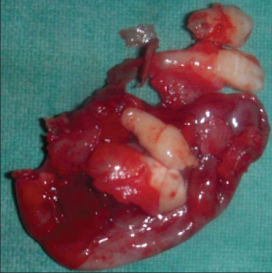 | Figure 5: Post-operative surgical specimen showing cystic lining along with the extracted teeth (Perman ent canine, supernumerary teeth, lateral incisor, retained deciduouscanine)
Click here to view |
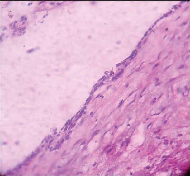 | Figure 6: Histopathological image of the cystic lining showing features of dentigerous cyst
Click here to view |
The lesion was found to be measuring around 3 cm × 3 cm, involving the sinus wall. The perforated sinus wall was repaired using autogenous bone graft from anterior sinus wall. Due precaution was taken to avoid perforation or tearing of lateral nasal wall, which was closely attached to the cystic lining. After complete removal of the cystic lining, the mucoperiosteal flap was approximated and suturing done using 3-0 Mersilk. Dressing was given. The post-operative healing was uneventful. The suture removal was carried out after 7 days. Histological examination of the specimen confirmed the diagnosis of a DC [Figure 6]. Patient was under follow-up for 6 months and presented no complications, meanwhile, he was advised for orthodontic and prosthetic rehabilitation to correct the malocclusion and alleviate problems with patients lack of dentition.
| Discussion | | |
Dentigerous cyst has been defined as one which is attached at the cement-enamel junction and encloses the crown of an unerupted tooth. Venous outflow, which is obstructed due to the pressure exerted by potentially erupting tooth on impacted follicle, induces rapid transudation of serum through capillary walls. The increased hydrostatic pressure of this pooling fluid separates the follicle from the crown with or without reduced enamel epithelium.
DC accounts for more than 24% of jaw cysts and is the most common developmental cyst of oral region. A very substantial majority involve mandibular third molar, maxillary permanent canine, followed by mandibular premolars and maxillary third molars in decreasing order. [6] Some of the studies have reported that dentigerous cyst arising from supernumerary teeth account for 5.29-7% of the cases. [3]
Patients with DC in maxillary bone or sinus may present with facial swelling, purulent rhinorrhea, nasal obstruction, external nasal deformity and epiphora. [7] Panoramic radiograph and upper occlusal radiograph are recommended as first-line diagnostic tools and further evaluation of the lesion by computed tomography examination. [8]
There is controversy regarding the management of large residual bony cavities with or without bone grafts. Numerous graft materials such as autografts, allogenic, xenografts and platelet rich plasma are available for the same. The surgical defect of the cystic cavity in our case was 3 cm × 3 cm. Relying on spontaneous bone regeneration no bone graft was used. The post-operative radiographs showed reduction in size of residual cavity and gradual increase in bone density. After 6 months, the healing and spontaneous filling of bone cavity was observed as with the studies of Shokier and Khalifa. [9] Long-term lack of dentition in a child may cause psychological or esthetic problems and also functional impairment.
The differential diagnosis of DC includes ameloblastoma, odontogenic keratocyst, odontogenic fibroma, odontogenic myxoma, cementomas and Pindborg tumor. [10] Early recognition of the entity and removal is necessary as they may rarely have the potential to develop odontogenic tumors like ameloblastoma and malignancy like squamous cell carcinoma and mucoepidermoid carcinoma. Esthetic and orthodontic problems are encountered when it is associated with anterior teeth. Hence, multidisciplinary management will help in better prognosis of these cases with restoration of esthetics and function.
| References | | |
| 1. | Ziccardi VB, Eggleston TI, Schneider RE. Using fenestration technique to treat a large dentigerous cyst. J Am Dent Assoc 1997;128:201-5. 
|
| 2. | Amin ZA, Amran M, Khairudin A. Removal of extensive maxillary dentigerous cyst via a Caldwell-Luc procedure. Arch Orofac Sci 2008;3:48-51.
|
| 3. | John T, Shekhar MG, Koshy M. Dentigerous cyst associated with supernumerary teeth: A report of three cases. J Clin Diagn Res 2010;4:2601-6.
|
| 4. | Dağistan S, Cakur B, Göregen M. A dentigerous cyst containing an ectopic canine tooth below the floor of the maxillary sinus: A case report. J Oral Sci 2007;49:249-52.
|
| 5. | McSherry PF. The ectopic maxillary canine: A review. Br J Orthod 1998;25:209-16.
[PUBMED] |
| 6. | Desai RS, Vanaki SS, Puranik RS, Tegginamani AS. Dentigerous cyst associated with permanent central incisor: A rare entity. J Indian Soc Pedod Prev Dent 2005;23:49-50.
[PUBMED]  |
| 7. | Onotai LO, da Lilly-Tariah OB. Dentigerous cyst associated with ectopic tooth at the roof of maxillary sinus. Int J Med Med Sci 2013;3:407-10.
|
| 8. | Jiang Q, Xu GZ, Yang C, Yu CQ, He DM, Zhang ZY. Dentigerous cysts associated with impacted supernumerary teeth in the anterior maxilla. Exp Ther Med 2011;2:805-9.
|
| 9. | Shokier HM, Khalifa GA. Assessment of bone healing in large bony defects after enucleation of jaw cysts without using any graft material using direct digital radiography and CT scan. C. D. J 2009;25:35-42.
|
| 10. | Ngamdu YB, Kodiya AM, Sandabe MB, Garandawa HI, Isa A. Dentigerous cyst associated with ectopic supernumerary canine in the maxillary sinus. J C R2012;2:27-30.
|
[Figure 1], [Figure 2], [Figure 3], [Figure 4], [Figure 5], [Figure 6]
|