|


 |
| CASE REPORT |
|
| Year : 2013 | Volume
: 5
| Issue : 2 | Page : 95-98 |
|
|
Total reconstruction of lower eyelid in a post-traumatic patient using modified fricke's cheek flap
Subhabrata Sengupta1, Binayak Baruah1, Suvra Pal2, Isha Preet Tuli1
1 Department of ENT, Sikkim Manipal Institute of Medical Sciences, Gangtok, Sikkim, India
2 Department of Eye, Sikkim Manipal Institute of Medical Sciences, Gangtok, Sikkim, India
| Date of Web Publication | 13-Mar-2014 |
Correspondence Address:
Subhabrata Sengupta
28/1B, Srimohan Lane, Kolkata - 700 026, West Bengal
India
 Source of Support: None, Conflict of Interest: None
DOI: 10.4103/2006-8808.128749

 Abstract Abstract | | |
Eyelids are very complex structure, reconstruction of which is a challenge to surgeons. Reconstruction of eyelids may be required in a variety of conditions like congenital anomalies, trauma, or postsurgical excision in malignant lesions involving the eyelids. There are numerous ways to reconstruct the eyelids; the best procedure depends on both the skill of the surgeon and the condition of the patient. Fricke' lateral temporal based flap was first described in 1829 for reconstruction of the eyelids and lateral canthal region. This flap had inherent problems regarding cosmetic appearance of the eyebrows. The modified Fricke's flap based on the cheek has the advantage of avoiding such complications. It is very easy and rapid outpatient department (OPD) based procedure with acceptable cosmetic and functional result. It can be done by all ear, nose, and throat (ENT) and head and neck surgeons without any reconstructive training. In this article we are presenting a case of total reconstruction of lower eyelid using the modified Fricke's cheek flap. Keywords: Lower eyelid reconstruction, modified Fricke′s cheek flap, post traumatic patient
How to cite this article:
Sengupta S, Baruah B, Pal S, Tuli IP. Total reconstruction of lower eyelid in a post-traumatic patient using modified fricke's cheek flap. J Surg Tech Case Report 2013;5:95-8 |
How to cite this URL:
Sengupta S, Baruah B, Pal S, Tuli IP. Total reconstruction of lower eyelid in a post-traumatic patient using modified fricke's cheek flap. J Surg Tech Case Report [serial online] 2013 [cited 2016 May 25];5:95-8. Available from: http://www.jstcr.org/text.asp?2013/5/2/95/128749 |
| Introduction | |  |
Like every other part of the human body, the eyelids are also a marvel in their own rights. Because of their shape and presence of beautiful eyelashes, the eyes appear as magnificent and expressive, and appeal to all human minds from time immemorial. Definitely it is a ghostly scenario to imagine the eyeball without having the eyelids. But apart from their cosmetic value, they have very important function to serve. They protect the eyes from trauma, excessive light, and also contribute in maintaining the biomechanics of the lacrimal apparatus, keeping the eyes moist. The architecture of the eyelids is very complex posing a real challenge to reconstruction. The aim, approach, and methods of reconstruction are different for upper and lower lids, as the functionality of the two lids differ. There are numerous methods of reconstruction of the eyelids. In this article, we describe a case of posttraumatic reconstruction of lower eyelid and periorbital area using a modification of the Fricke's flap.
| Case Report And Surgical Technique | | |
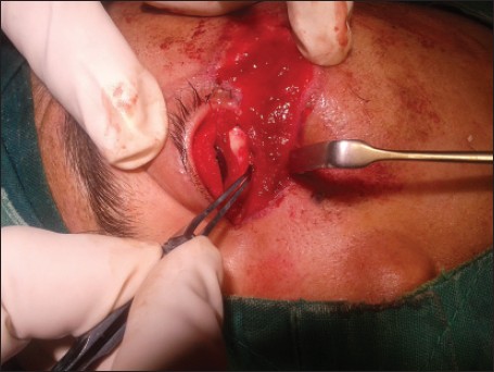
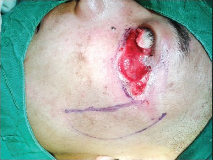
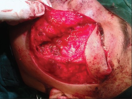
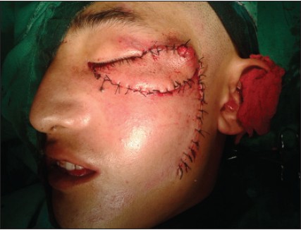
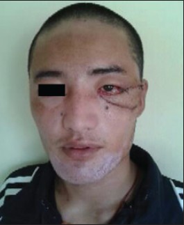
An 22-year-old male patient presented with avulsion injury of the left lower eyelid and lateral periorbital area following a road traffic accident. On examination it was revealed that there was complete loss of the lower eyelid including the skin, subcutaneous tissue, tarsal plate, and palpebral conjunctiva. Few fibers of the orbicularis oculi survived. There was extensive skin loss in the lateral periorbital area. There was no injury to the cornea, sclera, bulbar conjunctiva, lower fornix, and lacrimal apparatus. The upper eyelid was absolutely normal [Figure 1]. The patient was at first treated conservatively with antibiotics and regular dressing of the wound until fresh granulation tissue was formed. Then he was posted for reconstructive surgery with the aim of creating a functional lower eyelid with maximum possible cosmetic result. A strip of cartilage was harvested from the ear concha and inserted in a pocket created within the fibers of orbicularis to serve as tarsal plate [Figure 2]. The remnant of the conjunctiva at the lower fornix was sutured with the orbicularis fibers to form the posterior lamella. The anterior lamella was reconstructed using a modification of the Fricke's flap where the vertical limb of the flap was designed over the cheek. The entire flap design was sketched out on the skin starting from the wound margin with the vertical limb extending over the cheek [Figure 3]. It was slightly longer and wider than the defect to compensate for its retraction and included the totality of the subcutaneous cellular tissue. The length-to-width ratio was roughly 4:1. The whole flap was elevated according to the outline sketched over the skin, and thorough hemostasis was performed [Figure 4]. The apex between the donor and the receptor area is sutured first near the medial canthus; [Figure 5] followed by the margin with orbicularis oculi fibers, thus completing the anterior lamella. Rest of the defect over the cheek and the lateral temporal area was easily sutured primarily without any tension by adequate undermining of the margins resulting in satisfactory advancement displacement [Figure 6]. The repair was done in two layers. The subcutaneous layer was sutured with 5/0 vicryl and the skin margins opposed by horizontal mattress suture using 5/0 nylon except for the lid margin where simple suture was given avoiding excessive tension. The patient recovered smoothly without any significant postoperative complications. In the subsequent follow-up, the patient did not have major problems like irritation of the eyes, epiphora, synechia, or corneal ulcers [Figure 7].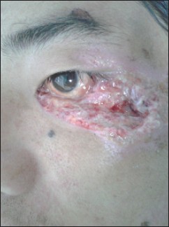 | Figure 1: Original trauma involving the skin, sub cutaneous tissue palpebral conjunctiva
Click here to view |
| Discussion | | |
Without basic knowledge of anatomy it is very difficult to conceptualize the reconstruction of eyelids. The lower lid is shorter in height, less mobile, and contributes minimally for palpebral closure. It has the thinnest skin in the body, which becomes thicker as it approaches the eyebrow and cheek. The skin is firmly adherent over the pretarsal area and over the medial and lateral canthal areas due to the absence of subcutaneous tissue. The anterior lamella consists of the skin and orbicularis muscle. The posterior lamella consists of the conjunctiva and tarsal plate. [1]
The main principle of eyelid reconstruction consists of three elements: An outer layer of skin; an inner layer of mucosa; and a semi rigid skeleton interposed between them. [2] One layer should carry its own blood supply and the other can be a free graft. The basic aim of reconstruction is to restore the anatomy and function of eyelids. Optimum results have to be achieved in terms of position, movements, and smooth internal lining capable of producing mucus for lubrication. Color match is important for cosmetic appearance of anterior lamella. Moreover in post injury cases, the obvious and anticipated tissue shortage must be precisely evaluated mainly over the forehead and cheek to prevent postoperative wound contracture. [1]
There are different techniques for total or partial lower eyelid reconstruction. In his case series on reconstruction of lower eyelids, Barba-Gómez et al., [3] have considered many such techniques like the method described by Mustarde, [2] the Hughes [4] transposition flap with its modifications, [5] the eyelid [6] cutaneous rim graft, the hard palate graft covered by an orbicularis oculi myocutaneous advancement flap, [7] the Tripier [8] flap, and more complex approaches such as the pre-expansion mucosa-lined tongue flap, [9] the use of acellular human dermis, [10],[11] the cheek flap supported by fascia lata, [12] the island tarsoconjuctival mucochondrocutaneous flap, [13],[14] and the use of an expanded forehead Fricke flap. [15],[16],[17] All of these techniques are useful when reconstruction of the lower eyelid is required; however, some of these procedures are complex and expensive.
The original Fricke flap is a temporally based monopedicle forehead transposition flap, first described by Johann Karl Fricke in 1829. [15] It can be used in the reconstruction of large lower lid, upper lid, and lateral canthal defects or to bring vascularized tissue to anterior orbital defects. [16] A laterally based skin flap above the eyebrow is raised and rotated to bring to the defect. The donor area is usually primarily closed or can be closed by skin graft. The flap revision is done in 2-week time. The disadvantage in Fricke's flap is raised eyebrow and scar as the eyebrow has to be pulled up for primary suturing.
The modified Fricke's cheek flap is a variation of this classical flap, as it is designed on the cheek rather than over the eyebrow. There are definite advantages of using this modification of the original flap designed by Fricke. Firstly, the cheek provides greater amount of tissue adjacent to the defect which matches in terms of color and texture. Secondly, it prevents elevation of the eyebrow and scarring. Thirdly, skin grafting can be avoided as the skin over the cheek can be easily primarily sutured with good cosmetic result. Fourthly, no flap revision is required after 2 weeks postoperatively. Finally, studies have shown that even without using chondromucosal graft, adequate mucosal regeneration leading to formation of a smooth and functional posterior lamella in this technique, minimizes the chances of corneal complications. The granulation tissue created at the bottom of the flap regenerates mucosa with time, which tends to contract and tether the flap downwards, thereby increasing scleral exposure. [3] The documented disadvantages include temporary chemosis of the conjunctiva coming in contact with the flap, scarring in the cheek, and some cases of corneal injury. Better cosmetic results can be achieved by additional interventions like flap lipectomy, scar revision, and eyelash implants. The posterior lamella can also be reconstructed with a periosteum flap to improve the results. [3] Keeping this in mind we have used the conchal cartilage with its perichondrium as an attempt to improve the functional and cosmetic result in our patient. Being a vascularized flap, it can support a free graft for the reconstruction of the posterior lamella.
There are definite advantages of the modified Fricke's flap in reconstruction of lower eyelid over other techniques described in the literature. Firstly, it is useful in the monocular patient in whom occlusion of the visual axis cannot be done as in techniques such as the Hughes or Cutler-Beard flaps. Secondly, it can be used for the reconstruction of an extensive lower eyelid defect, which is too large for a Hughes cutler flap. Thirdly, extensive dissection involved in a Mustarde cheek rotation flap with the attendant risk of facial nerve trauma can be avoided. It is particularly useful in lower lid defects involving the entire length of the lower lid, but which are relatively short in their vertical dimension. [16]
| Conclusion | | |
Of the various techniques available for the reconstruction of lower eyelid, one has to choose the procedure which suits the surgeon and the patient both. Consideration has to be given on the nature of the lesion, area of involvement, degree of tissue loss, condition of the patient, and above all the skill of the surgeon and availability of plastic surgery colleagues. Keeping in mind that the lower eyelid has minimal contribution to eye closure, rather its main function is to render stability, the end result of the reconstructive procedure must make the reconstructed eyelid more stable. The issue of cosmetic and esthetic appearance is also of profound importance. The modified Fricke's flap based on the cheek is a very simple technique for reconstruction of lower eyelid. It has the advantage of avoiding the complications of the original Fricke's flap technique. It can be easily done by all head and neck surgeons without any special reconstructive training. The result may not be excellent in terms of cosmetic appearance, but certainly not unacceptable. It is able to provide a functional lower eyelid to a person in whom it was lost due to ailment or trauma. Moreover, this simple technique is a rapid, inexpensive one which can be done on outpatient department (OPD) basis not requiring any special setup. It has very low complication rate and low patient morbidity. Although advocated by some surgeons as a 'last-resort' [18] method of periorbital reconstruction, it is a valuable option with acceptable cosmetic and functional results.
| References | | |
| 1. | Subramanian N. Reconstruction of eyelid defects. Indian J Plast Surg 2011;44:5-13. 
[PUBMED]  |
| 2. | Mustarde JC. Repair and reconstruction in the orbital region. 2 nd ed. Philadelphia: Churchill Livingstone; 1991.
|
| 3. | Barba-Gómez J, Zuñiga-Mendoza O, Iñiguez-Briseño I, Sánchez-Tadeo MT, Barba-Gómez JF, Molina-Frechero N, et al. Total lower-eyelid reconstruction: Modified Fricke's cheek flap. J Plast Reconstr Aesthet Surg 2011;64:1430-5.
|
| 4. | Hughes WL. Total lower eyelid reconstruction: Technical details. Trans Am Ophthalmol Soc 1976;74:321-9.
[PUBMED] |
| 5. | Rohrich RJ, Zbar RI. The evolution of the Hughes tarsoconjunctival flap for the lower eyelid reconstruction. Plas Reconstruc Surg 1999;104:518-22.
|
| 6. | O'Donnell BA. The cutaneomarginal eyelid graft. Clin Experiment Ophthalmol 2002;30:136-9.
[PUBMED] |
| 7. | Lalonde DH, Osei-Tutu KB. Functional reconstruction of unilateral, subtotal, full-thickness upper and lower eyelid defects with a single hard palate graft covered with advancement orbicularis myocutaneous flaps. Plast Reconstr Sur 2005;115:1696-700.
|
| 8. | Herman AR, Bennet RG. Reconstruction of a large surgical defect on the lower eyelid and infraorbital cheek. Dermatol Surg 2005;31:689-91.
|
| 9. | Miyawaki T, Hisako A, Suzuki H, Kurihara K, Jackson IT. Pre-expansion of mucosa lined flap for lower eyelid reconstruction. Plast Reconstr Surg 2005;116:76e-82e.
|
| 10. | Li TG, Shorr N, Goldberg RA. Comparison of the efficacy of hard palate grafts with acellular human dermis grafts in lower eyelid surgery. Plast Reconstr Surg 2005;116:873-8.
|
| 11. | Taban M, Douglas R, Li T, Goldberg RA, Shorr N. Efficacy of "thick" acellular human dermis (AlloDerm) for lower eyelid reconstruction: Comparison with hard palate and thin alloderm grafts. Arch Facial Plast Surg 2005;7:38-44.
|
| 12. | Matsumoto K, Nakanishi H, Urano Y, Kubo Y, Nagae H. Lower eyelid reconstruction with a cheek flap supported by fascia lata. Plast Reconstr Surg 1999;103:1650-4.
|
| 13. | Porfiris E, Christopoulos A, Sandris P, Georgiou P, Ioannidis A, Popa CV, et al. Upper eyelid orbicularis oculi flap with tarsoconjunctival island for reconstruction of full-thickness lower lid defects. Plast Reconstr Surg 1999;103:186-91.
|
| 14. | Porfiris E, Georgiou P, Harkiolakis G, Popa CV, Sandris P, Sgouras N. Island mucochrondrocutaneous flap for reconstruction of total loss of the lower eyelid. Plast Reconstr Surg 1997;100:104-7.
|
| 15. | Fricke JC. Die Bildung neuer Augenlider (Blepharoplastik) nach Zersto¨rungen und dadurch hervorgebrachten Auswa ¨rtswendungen derselben. Hamburg: Pethes und Bessler; 1829.
|
| 16. | Wilcsek G, Leatherbarrow B, Halliwell M, Francis I. The 'RITE' use of the Fricke flap in periorbital reconstruction. Eye 2005;19:854-60.
|
| 17. | Salomon J, Bieniek A, Baran E, Szepietowski JC. Basal cell carcinoma on the eyelids: Own experience. Dermatol Surg 2004;30:257-63.
|
| 18. | McCord D. Upper eyelid reconstruction. In: McCord CD, editor. Eyelid surgery. Principles and techniques. 1 st ed. Philadelphia: Lippincott-Raven; 1995. p. 252-69.
|
[Figure 1], [Figure 2], [Figure 3], [Figure 4], [Figure 5], [Figure 6], [Figure 7]
|