|
 
 |
| ORIGINAL ARTICLE |
|
| Year : 2017 | Volume
: 5
| Issue : 1 | Page : 14-17 |
|
Sexual dimorphism in frontal sinus of Southeast Nigerians
Uchenna Kenneth Ezemagu1, C I. P Anibeze2, Frank Chinedu Akpuaka3
1 Department of Anatomy, Federal University, Ndufu-Alike Ikwo, Ebonyi, Nigeria
2 Department of Anatomy, College of Medicine, Enugu State University, Enugu, Nigeria
3 Department of Anatomy, Abia State University, Uturu, Nigeria
| Date of Web Publication | 11-Jul-2017 |
Correspondence Address:
Uchenna Kenneth Ezemagu
Department of Anatomy, Federal University, Ndufu-Alike Ikwo, Ebonyi
Nigeria
 Source of Support: None, Conflict of Interest: None  | Check |
DOI: 10.4103/2315-7992.210248

Background: The functional and adaptive significance of frontal sinus is still very poorly understood and attempt to link frontal sinus patterns with specific influences such as sex or biological affinity have been inconclusive. Purpose: The study aims to demonstrate the influence and this relationship between the variation in frontal sinus dimensions and sex. Materials and Method: In this study, lateral and anteroposterior radiographs of 74 males and 46 females of Southeast Nigerians taken from August 18, 2003 to July 12, 2004 were measured (age 9–75). Data were analyzed using excel package of a desktop computer, employing Chi-square test to determine the association between sinus dimensions and sex. Result: The result showed that variation in frontal sinus breadth and depth may not depend on sex while variation in height slightly depends on sex (P < 0.05). Thus, establishing the fact that some metric and morphologic characteristics of the frontal sinus depend on sex. Conclusion: This finding depicts a sexual dimorphism in frontal sinus dimension. Moreover, it adds an additional factor to the puzzle of the meaning of the supra orbital development and morphologic characteristics. Keywords: Dimorphism, frontal sinus, pneumatization, sex
How to cite this article:
Ezemagu UK, Anibeze C I, Akpuaka FC. Sexual dimorphism in frontal sinus of Southeast Nigerians. Ann Bioanthropol 2017;5:14-7 |
| Introduction | |  |
The extent of frontal sinuses varies among individuals and between the two sides. The right and left frontal sinuses are rarely of equal size and the septum between the right and left sinuses usually is not situated in the median plane.[1] Moreover, often a frontal sinus has two parts: A vertical part in the squamous part of the frontal bone and a horizontal part in the orbital part of the frontal bone. In addition, the frontal sinus may extend into the crista galli.[2] The frontal sinuses are connected to the middle meatus via the frontal recess, which allows secretions to drain.[3] Variation in frontal sinus has been suggested to be attributable to sex [4] and biological affinity.[2] The dimensions of the frontal sinus are higher for males than females with mean length and width of frontal sinuses as 35.36 mm, 63.87 mm for males and 28.70 mm, 60.73 mm for females, respectively, in a population of southeast Nigerians.[2]
The biochemical regimes associated with diet that differ in occlusal loading may have an effect on paranasal sinus size, although the relationship between masticatory stress and pueumatization is unclear at present.[5] However, no one theory or explanation for variation has emerged as dominant.[2] This ambiguity may be due in part to the lack of understanding of the phylogenetic distribution of sinuses across taxa.[6] The size and shape of maxillary sinus determine to a considerable extent the facial appearance such that a small narrow sinus with a concave anterior wall gives rise to a dish face and a large sinus with a convex anterior wall gives rise to a round face.[7] Moreover, the anterior wall of each frontal sinus is responsible for the prominence of the forehead, which is situated above the eye brow, but the shape of the supra orbital ridges and the forehead are no indication of their size.
The importance of understanding variation in fontal sinus lies on two aspects. First, their pathological conditions may be understood. Second, researchers in paleontology [8] have offered a variety of hypotheses concerning development of the supra orbital region. If it can be shown that supraorbital development is even partially correlated with sinus size and sex, it adds an additional factor to the puzzle of the meaning of supraorbital development.
There is a paucity of literature on the variations of the sinuses in Nigeria or Africa in general. Anthropologists were linking sinus morphology and development to races and evolution.[9] The aim of this study was to observe the sexual dimorphism in the frontal sinuses of Southeast Nigerians.
| Materials and Methods | | |
A total of 120 radiographs (74 males and 46 females) with age ranging from 9 to 75 years with lateral and Caldwell (anteroposterior) views were measured at Ebonyi State Teaching Hospital, Abakaliki, University of Nigeria Teaching Hospital Enugu and Hansa Clinic, Enugu. Although the samples of radiographs used were collected from different places, the focus film distance used was the same (90 cm).
Radiographs of sinus used include those reported normal by the radiologists and those without cases of previous sinus operation.
The three parameters measured were height on the vertical part, breadth, and depth on the horizontal part of frontal sinuses. The height and depth of the frontal sinuses were taken from the lateral view radiographs. Frontal sinus mostly appears as a shadow with its apex superiorly and base/floor inferiorly shown. The height was measured in vertical plane from the (A) apex to the base (B) as shown in [Figure 1]. The depth or anteroposterior dimension was taken as the longest line perpendicular to the length and touching the (C) anterior and (D) posterior tables of the frontal sinus at its floor using the protractor and metric rule, because of its irregularly shaped floor.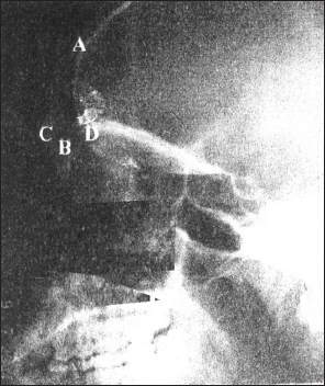 | Figure 1: A plate showing land-marks used for measurement of Length (AB), Depth (CD) of frontal sinus. A = Apex of Sinus. B = Roof of Orbit C = Anterior Table D = Posterior Table
Click here to view |
The breadth of the frontal sinus was taken from the anteroposterior view radiographs. It appears as a crescent shaped shadow in this view separated in to two or three lobes by thin septa. Because of difficulty delimiting the inferior borders of the frontal sinus previous researchers have often drawn a base line (CD) tangential to the superior borders of the orbit as shown in [Figure 2].[2],[10] The breadth was taken as the longest (AB) line parallel to the baseline touching the left and right lateral edges of the frontal sinus using the sets of squares and metric rule. In cases of smaller frontal sinuses, which may be partially or completely eliminated in these procedures, the parallel line was taken midway between the baseline and the nasion (the midpoint of the nasofrontal suture).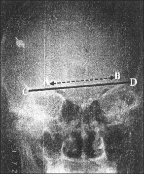 | Figure 2: A plate showing land-marks for measurement of width /AB/ of frontal sinus. A = left lateral edge of frontal Sinus. B = Right lateral edge of frontal, /CD/ = Baseline
Click here to view |
Statistical analysis
The sex, height, breadth, and depth of the frontal sinuses were condensed in groups. Cross tabulations were done on these groups for sex. This continued with Chi-square test designed to test the degree of association between height, breadth, and depth of the sinus by sex. When the null hypothesis of no association was rejected at α = 0.05 (95% confidence), an association was established between the two variables under analysis, and the degree of dependency was calculated.
| Results | | |
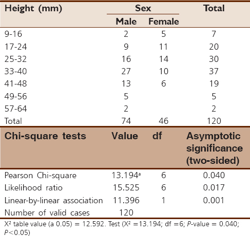
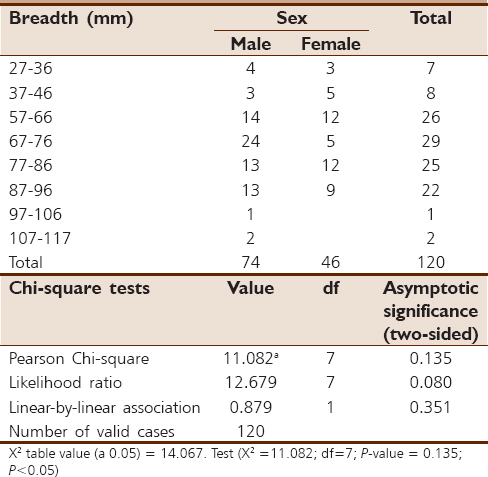
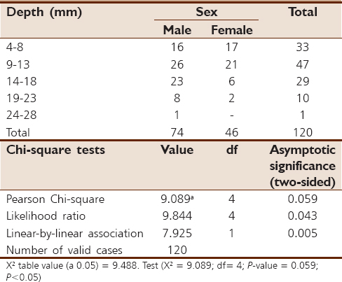
The relationship between sex and height of the frontal sinus was shown in [Table 1]. The data showed that the null hypothesis of no association should be rejected at α = 0.05. The degree of dependency showed that 31% of the variation in frontal sinus height was dependent on variation in sex. The relationship between sex and breadth or depth as shown in [Table 2] and [Table 3] is indicative of the fact that the evidence to reject the null hypothesis of no association was lacked at α = 0.05.
| Discussion | | |
The study showed that some metric and morphologic characteristics of frontal sinus depend on sex. Variation in the frontal sinus may be attributable to sex.[4] The value for the dimensions of frontal sinus are higher for males than females [2] although the degree of dependency in this study showed that only 31% of the variation in frontal sinus height was dependent on variation in sex demonstrating a positive relationship.
The relationship between sex and frontal sinus breadth and depth showed that the evidence to reject the null hypothesis of no association was lacked. It therefore follows that frontal sinus breadth and depth may not vary with sex, which suggests no sexual dimorphism in the horizontal portion of the frontal sinus. The observed sexual dimorphism could not be explained since possible epigenetic determinants for sizes of paranasal sinuses such as temperature, masticatory stress,[5] shapes and size of forehead,[7] some metric and morphologic characteristics of supraorbital region were not dealt with in this study.
| Conclusion | | |
This finding depicts a sexual dimorphism in frontal sinus dimension. Moreover, it adds an additional factor to the puzzle of the meaning of the supraorbital development and as such may bring us closer to understanding the basis of variation in the supraorbital region of modern Homo sapiens. Clearly, additional tests are necessary to explore the role played by genetic and epigenetic factors in the morphology of the paranasal sinuses.
Acknowledgment
The authors thank P.J. Eze for advice and support, Hansa Clinic for access to the radiograph facilities and Felix Aguboshim for helping in the statistical analysis of this work.
Financial support and sponsorship
Nil.
Conflicts of interest
There are no conflicts of interest.
| References | | |
| 1. | Keith MI, Arthur DI. Clinically Oriented Anatomy. 4 th ed. Philadelphia: Lippincott Williams and Wilkins; 1999. p. 961.  |
| 2. | Ezemagu UK, Mgbor N, Anibeze CI, Akpuaka FC. Anthropometrical profiles of frontal sinus in population of Southeast Nigerians. J Exp Clin Anat 2005;4:42-6. |
| 3. | Ezemagu UK, Mgbor N, Anibeze CI, Akpuaka FC. Anthropometrical profiles of maxillary sinus in population of Southeast Nigerians. J Biomed Sci Afr 2010;8:15-9. |
| 4. | Buckland-Wright JC. A radiographic examination of frontal sinuses in early British populations. Man 1970;5:512-7. |
| 5. | Rae TC, Hill RA, Hamada Y, Koppe T. Clinal variation of maxillary sinus volume in Japanese macaques ( Macaca fuscata). Am J Primatol 2003;59:153-8. [ PUBMED] |
| 6. | Rae TC, Koppe T, Spoor F, Benefit B, McCrossin M. Ancestral loss of the maxillary sinus in old world monkeys and independent acquisition in Macaca. Am J Phys Anthropol 2002;117:293-6. [ PUBMED] |
| 7. | Romanes GJ. Cunningham's Textbook of Anatomy. 12 th ed. New York: Oxford University Press; 1981. p. 124-5. |
| 8. | Oyen OJ, Rice RW, Cannon SM. Browridge structure and function in extant primates and Neanderthals. Am J Phys Anthropol 1979;51:83-96. |
| 9. | Frayer DW. Evaluation at the European edge: Neanderthal and upper paleolithic relationships. Prehist Europ 1992;2:9-69. |
| 10. | Francis P, Raman R, Korula P, Korah I. Pneumatization of the paranasal sinuses (maxillary and frontal) in cleft lip and palate. Arch Otolaryngol Head Neck Surg 1990;116:920-2. [ PUBMED] |
[Figure 1], [Figure 2]
[Table 1], [Table 2], [Table 3]
|


 Search Pubmed for