|
 
 |
|
CASE REPORT |
|
|
|
| Year : 2012 | Volume
: 18
| Issue : 1 | Page : 127-129 |
| |
Dysmorphic features and congenital heart disease in chromosome 6q deletion: A short report
Sreelata Nair, Rini Varghese, Sajeed Hashim, Pappachan Scariah
Department of Fetal Medicine, Lifeline Genetics and Research Centre, Lifeline Super Specialty Hospital, Adoor, Pathanamthitta, Kerala, India
| Date of Web Publication | 26-May-2012 |
Correspondence Address:
Sreelata Nair
Department of Fetal Medicine, Lifeline Genetics and Research Centre, Lifeline Super specialty Hospital, Adoor, Pathanmthitta, Kerala - 691 523
India
 Source of Support: None, Conflict of Interest: None  | 4 |
DOI: 10.4103/0971-6866.96682

 Abstract Abstract | | |
In this report, we describe a one and a half year old girl showing terminal deletion of long arm of chromosome 6q. The associated abnormalities such as congenital heart disease, mental retardation, and dysmorphic features are described. Cytogenetic studies with GTG banding showed 46,XX,del(6)(q24→qter). Karyotype of the parents was normal suggesting a denovo event.
Keywords: Chromosome, deletion, dysmorphism, genetic counseling, karyotype
How to cite this article:
Nair S, Varghese R, Hashim S, Scariah P. Dysmorphic features and congenital heart disease in chromosome 6q deletion: A short report. Indian J Hum Genet 2012;18:127-9 |
How to cite this URL:
Nair S, Varghese R, Hashim S, Scariah P. Dysmorphic features and congenital heart disease in chromosome 6q deletion: A short report. Indian J Hum Genet [serial online] 2012 [cited 2016 Jun 1];18:127-9. Available from: http://www.ijhg.com/text.asp?2012/18/1/127/96682 |
| Introduction | |  |
Chromosomal deletions may arise denovo as well as from unbalanced translocations. Denovo deletions may affect the terminal part of a chromosome or an interstitial region, and usually affect a fairly large number of genes and produce recognizable syndromes. Microscopically visible deletions generally involve multiple genes, and the consequences of losing this much genetic material from even one of the chromosome pair can be severe. [1] If the missing part contains important instructions, it can lead to congenital malformations, development delay, or mental retardation.
| Case Report | | |
A one and a half year old girl child born of non-consanguineous marriage to a 27-year-old primi mother was referred to the genetic clinic. The child was born at term by elective cesarean section because of symmetric IUGR and oligohydramnios. Birth weight was 2.0 kg and length was 48 cm with head circumference 30 cm. The baby was admitted to NICU in view of respiratory distress soon after birth. There was no significant family history, no evidence of teratogenecity, and no other pregnancy associated complications.
Clinical examination of the new born revealed grade 3 pansystolic murmurs in the left lower sternal border. ECHO was suggestive of congenital acyanotic heart disease VSD, ASD, PDA with moderately severe pulmonary hypertension. USG abdomen was normal.
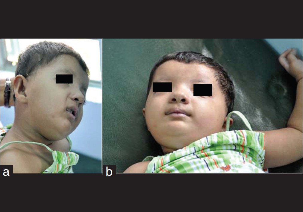
The first genetic consultation was at the age of 1 year 6 months for dysmorphic features and congenital heart disease. Facial dysmorphism included microcephaly, brachycephaly, hypertelorism, upslanting palpebral fissures, strabismus, broad nasal bridge, wide anterior fontanalle, anteverted nares, high arched palate, microretrognathia, low set dysmorphic ears, and short neck. Clitoromegaly with prominent labia minora was seen. Congenital acyanotic heart disease with vsd,0 ASD, and pda0 was noted. Pallor was seen along with cyanosis and nail bed clubbing of the finger and toes. Psychomotor development was extremely delayed and marked hypotonia was seen. MRI (brain) showed partial agenesis of corpus callosum, bilateral mildly enlarged subarchanoid space, and mildly dilated third and lateral ventricles.
Cytogenetic findings
Cytogenetic analysis from the proband was carried out by peripheral blood lymphocyte culture [2] and GTG banding. Twenty metaphase were analyzed after GTG banding at 500 band resolution. Karyotype of the proband showed deletion in the terminal region of the long arm of chromosome 6 in all the metaphase analyzed. The karyotype of the child was 46,XX,del(6)(q24→qter) [Figure 1]a and b.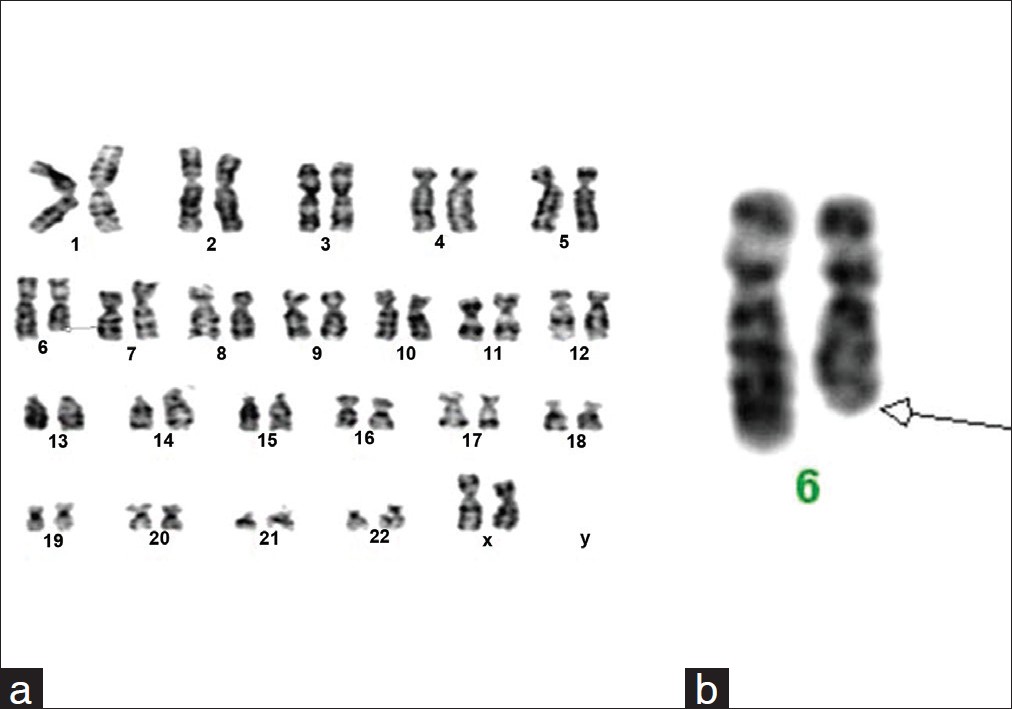 | Figure 1: (a) Karyotype 46,XX,del(6)(q24 to qter) showing deletion, (b) Partial karyotype showing 6q 24 to terminal deletion
Click here to view |
| Discussion | | |
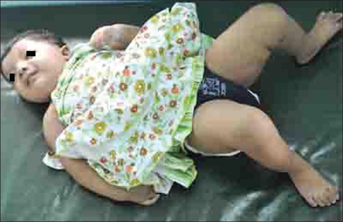
Here, we describe a case with deletion in the terminal region of long arm of chromosome 6 with break point at q 24 region. Chromosome 6q deletion is associated with dysmorphic features and multiple anomalies, which were seen in the index case also. The more frequently observed clinical features in 6q deletion are congenital heart defects, [3] which were also seen in our case. The child had VSD, ASD, and PDA with moderately severe pulmonary hypertension. Other features like brachydactyly, clinodactyly along with peripheral clubbing and cyanosis were also seen [Figure 2]a, b and [Figure 3]. Literature search with chromosome 6q24 deletion showed few reports with varying other anomalies like nuchal cyst, diaphragmatic hernia, multicystic kidney, etc, which was not seen in our case. [4],[5] Caselli et al., [6] have reported a similar case with deletion of 6q24.3-25.1 in an 8-year-old with growth failure, cardiac septal defects, mental retardation, and dysmorphic features.
The region involved in our case is q24 to terminal and therefore explains the neurodevelopmental delay and peculiar facial features. This critical region has a 2.6 Mb deletion. More than twenty two genes are present in this deleted region, [6] which might be important in the normal development of cardiovascular and central nervous system. The karyotype of parents were normal, which further suggests that this must be a denovo event and not due to unbalanced product of familial translocation. As the parental karyotype was normal and the child had a denovo deletion, the chance of recurrence is very low. Nevertheless, prenatal diagnosis by amniocentesis and fetal karyotyping has been offered to the couple, in case of future pregnancies. Genetic counseling was given in detail.
Hence, chromosome analysis should be offered to every child with mental retardation and dysmorphism as in this case. FISH studies and array-CGH analysis may be useful to delineate the size of the deletion and the genes involved, which in our case was not affordable by the patient.
| References | | |
| 1. | Jorde LB, Carey JC, Bamshad MJ. Chromosomal Basis of Human Disease. In: Medical Genetics. 4 th ed. Haryana: Elsevier Publishers; 2010. p. 100-27. 
|
| 2. | Hungerford DA. Leukocyte cultured from small inocula to whole blood and the preparation of metaphase chromosome by treatment with hypotonic KCl. Stain Technol 1965;40:333-5.
[PUBMED] |
| 3. | Boy R, Pimentel MG, Hemerly AP, Silva MP, Barreiro AP, Almeida JC, et al. Chromosome 6q deletion: Report of a new case and review of the literature. Genet Mol Biol 1998;21:1415-4757.
|
| 4. | Krassikoff N, Sekhon GS. Terminal deletion of 6q and Fryns syndrome. A micro deletion/syndrome pair. Am J Med Genet 1992;36:363-4.
|
| 5. | Valtat C, Galliano D, Mettey R, Toutain A, Moraine C. Monosomy 6q: Report on four new cases. Clin Genet 1992;41:159-66.
[PUBMED] |
| 6. | Caselli R, Mencarelli MA, Papa FT, Uliana V, Schiavone S, Strambi M, et al. A 2.6 Mb deletion of 6q 24.3-25.1 in a patient with growth failure, cardiac septal defect, thin upper lip and asymmetric dysmorphic ears. Eur J Med Genet 2007;50:315-21.
|
[Figure 1], [Figure 2], [Figure 3]
| This article has been cited by | | 1 |
F8 genetic analysis strategies when standard approaches fail |
|
| B. Pezeshkpoor,A. Pavlova,J. Oldenburg,O. El-Maarri | | Hämostaseologie. 2013; 34(2) | | [Pubmed] | [DOI] | |
|
|
|
|