|
 
 |
|
BRIEF REPORT |
|
|
|
| Year : 2012 | Volume
: 18
| Issue : 3 | Page : 344-345 |
| |
Rhizomelic chondrodysplasia punctata: A missed opportunity for early diagnosis
Nanda Chhavi, Sankar Prashanth, Chandrasekaran Venkatesh, Kadirvel Karthikeyan
Department of Pediatrics, Mahatma Gandhi Medical College and Research Institute, Pillaiyarkuppam, Puducherry, India
| Date of Web Publication | 4-Mar-2013 |
Correspondence Address:
Chandrasekaran Venkatesh
Assistant Professor of Pediatrics, Mahatma Gandhi Medical College and Research Institute, Pillaiyarkuppam, Puducherry - 607 402
India
 Source of Support: None, Conflict of Interest: None
DOI: 10.4103/0971-6866.107990

 Abstract Abstract | | |
A male neonate was born with rhizomelic shortening of limbs. Skeletal radiograph showed punctate calcification of epiphysis of humerus, femur, and tibia. The diagnosis and a brief review of literature pertaining to the condition with emphasis on antenatal diagnosis and counseling are being reported.
Keywords: Counseling, prenatal diagnosis, rhizomelic chondrodysplasia punctata
How to cite this article:
Chhavi N, Prashanth S, Venkatesh C, Karthikeyan K. Rhizomelic chondrodysplasia punctata: A missed opportunity for early diagnosis. Indian J Hum Genet 2012;18:344-5 |
How to cite this URL:
Chhavi N, Prashanth S, Venkatesh C, Karthikeyan K. Rhizomelic chondrodysplasia punctata: A missed opportunity for early diagnosis. Indian J Hum Genet [serial online] 2012 [cited 2016 Jun 1];18:344-5. Available from: http://www.ijhg.com/text.asp?2012/18/3/344/107990 |
| Introduction | |  |
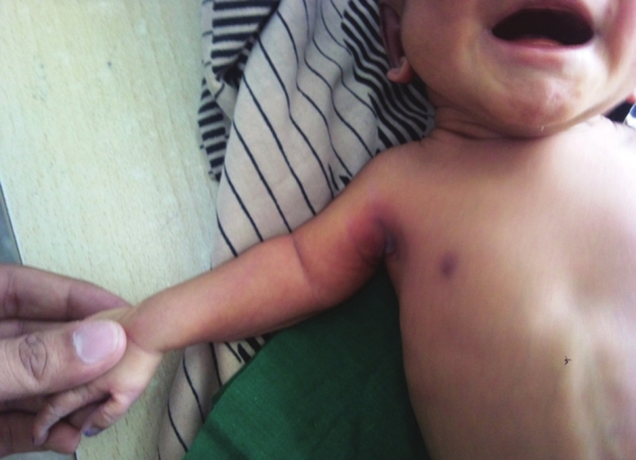
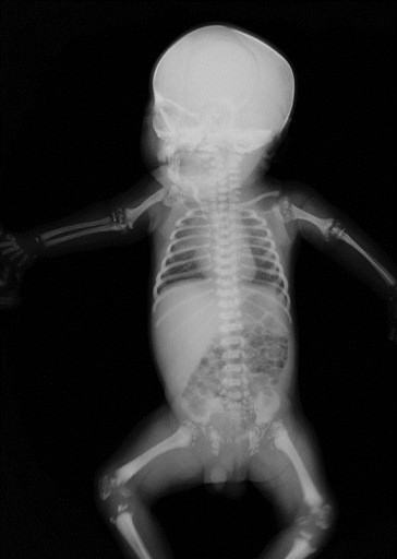
A male neonate born to G2 P1L1 mother at term by spontaneous vaginal delivery to III degree consanguineous marriage was found to have proximal shortening of both upper and lower limbs [Figure 1]. The antenatal period was uneventful and antenatal ultrasound was reportedly not done during pregnancy and the mother was referred to our hospital after the onset of labor. There was no family history of similar births and parents were phenotypically normal. Apart from rhizomelic shortening, the neonate also had coronal clefts of thoracic vertebrae and stippled epiphysis of femur tibia and humerus on skeletal survey radiograph [Figure 2]. There were no other congenital anomalies or dysmorphic facies. Based on the above features a provisional diagnosis of Rhizomelic Chondro-dysplasia Punctata (RCDP) was made and the prognosis was explained to the parents. The baby developed progressively severe respiratory distress and was discharged at request on day 3 of life as the parents were unable to come to terms with the diagnosis.
Chondrodysplasia punctata is a radiological diagnosis characterized by punctate or stippled calcifications in epiphyseal cartilage and seen in peroxisomal disorders such as Zellweger syndrome, neonatal adrenoleukodystrophy, and infantile Refsum disease. It may also be inherited as X-linked dominant, X-linked recessive, and autosomal recessive forms. [1] RCDP is a single gene defect leading to decreased plasmalogen synthesis. It is classically associated with PEX7 gene (peroxin family of genes) mutation [2] and has been reported in Indian patients too. [3] RCDP is characterized by proximal shortening of the humerus and to a lesser degree the femur, punctate calcifications in cartilage with epiphyseal and metaphyseal abnormalities, radiolucent defects (coronal clefts) of the vertebral bodies which represents cartilage that are not ossified, cataracts, contractures, microcephaly, characteristic skin changes of icthyosis, facial dysmorphism (depressed nasal bridge, hypertelorism, hypoplastic midface, anteverted nostrils, full cheeks), and developmental impairment.
This condition is considered to be lethal and most of the affected fetuses die in utero or soon after birth. Only few of them survive beyond infancy with severe physical disability and profound mental retardation in whom, death usually occurs in the first decade of life. Diagnosis of RCDP is based on clinical findings and confirmed by clinically available biochemical or molecular genetic testing which includes biochemical tests of peroxisomal function like red cell plasmologen concentration, plasma phytanic acid, and very long chain fatty acid estimation. [4] Sonological diagnosis can be reliably made between 19 and 21 weeks of gestation. [5]
This case is presented due to its rarity and failure to detect such an abnormality in utero resulting in a wasted pregnancy. The lack of resources (both money and manpower) is probably responsible for this tragedy to the parents which could have been prevented by early diagnosis and appropriate counseling. Establishing regional genetic labs which are connected with district level hospitals can be of immense help in reducing the burden of genetic diseases by appropriate prenatal diagnosis and counseling.
| References | | |
| 1. | Irving MD, Chitty LS, Mansour S, Hall CM. Chondrodysplasia punctata: A clinical diagnostic and radiological review. Clin Dysmorphol 2008;17:229-41. 
[PUBMED] |
| 2. | Braverman N, Steel G, Obie C, Moser A, Moser H, Gould SJ, et al. Human PEX7 encodes the peroxisomal PTS2 receptor and is responsible for rhizomelic chondrodysplasia punctata. Nat Genet 1997;15:369-76.
[PUBMED] |
| 3. | Phadke SR, Gupta N, Girisha KM, Kabra M, Maeda M, Vidal E, et al. Rhizomelic chondrodysplasia punctata type 1: Report of mutations in 3 children from India. J Appl Genet 2010;51:107-10.
[PUBMED] |
| 4. | Braverman NE, Moser AB, Steinberg SJ. Rhizomelic Chondrodysplasia Punctata Type 1. In: Pagon RA, Bird TD, Dolan CR, Stephens K, editors. Gene Reviews [internet]. Seattle (WA): University of Washington, Seattle; 1993-2001.
[PUBMED] |
| 5. | Schramm T, Gloning KP, Minderer S, Daumer-Haas C, Hortnagel K, Nerlich A, et al. Prenatal sonographic diagnosis of skeletal dysplasias. Ultrasound Obstet Gynecol 2009;34:160-70.
|
[Figure 1], [Figure 2]
|